
MIT Microscope Captures Millisecond-Scale Voltage Activity Across Zebrafish Brains
MIT researchers adapted a light-sheet microscope to image electrical activity across the brains of larval zebrafish at up to 200 scans per second. The method captured individual voltage spikes and brain-wide responses to ultraviolet light, although usable signals appeared in about one-quarter of neurons and the system remains an experimental research tool.
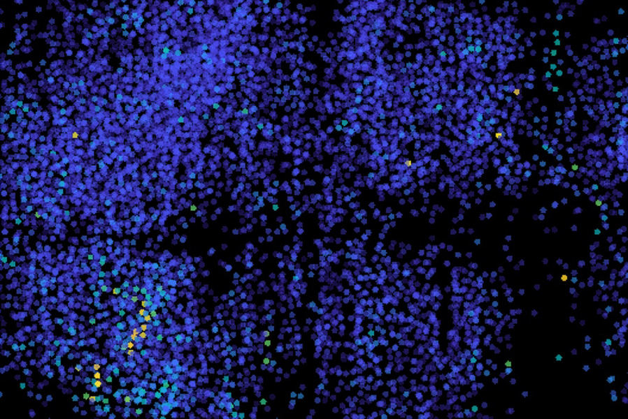
MIT researchers have developed a microscope-based method that can track electrical activity across the brain of a living larval zebrafish at millisecond-scale resolution. The approach, described in a study published August 14 in Nature Methods, is designed to help researchers examine how distributed groups of neurons work together rather than focusing on a small region at a time.
The team modified a light-sheet microscope, which uses a thin sheet of laser light to image successive layers of a sample and build three-dimensional views. To make whole-brain imaging fast enough to capture individual electrical impulses, the researchers increased camera acquisition speed and used remote refocusing to accelerate scanning.
With those changes, the microscope could scan an entire zebrafish brain 200 times per second, or once every five milliseconds. That speed allowed the researchers to observe voltage spikes and rapid bursts of activity from neurons distributed throughout the brain.
The system relies on genetically encoded voltage indicators rather than conventional calcium imaging. Calcium imaging measures the influx of calcium that follows neuronal firing, but the MIT researchers said it is generally too slow to resolve individual spikes. Voltage indicators are fluorescent proteins that respond directly to changes in a neuron's electrical state. For the experiments, the researchers engineered larval zebrafish neurons to express an indicator called Positron2-Kv.
The indicator did not produce usable signals in every neuron. About one-quarter of the neurons showed acceptable signals, but that coverage was sufficient for the team to identify brain-wide activity patterns. In resting fish, the researchers observed individual voltage spikes as well as rapid sequences of spikes.
They also examined how neural activity changed after exposure to ultraviolet light. Activity appeared first in the optic tectum, a brain region that receives and processes visual information from the retina. The activity then spread from one side of the tectum to the other. The researchers also observed stimulus-independent sequences involving groups of neurons in the cerebellum and hindbrain.
The study’s findings come from an experimental zebrafish model, not from human participants. The technique therefore demonstrates a research capability rather than establishing how human brains generate behavior, emotions, or mental states. The researchers said they hope to improve the proportion of neurons that can be measured, along with the microscope’s speed and resolution, and to extend the method to other experimental models, including mice.
The team’s broader aim is to give neuroscientists a way to generate hypotheses about how neural networks operate during specific behaviors and brain states. The method may be particularly useful for studying activity that unfolds across multiple brain regions at the same time, a pattern that slower or more localized imaging approaches may not capture.
Reporting Note
Biohack Report distinguishes preliminary findings, clinical evidence and commercial claims whenever the available reporting supports that distinction. Coverage is informational and is not medical advice.