
Injectable Scaffold Promotes Stroke Recovery in Mice by Recruiting Immune Cells
A Duke University study found that an injectable hydrogel scaffold helped mouse brains damaged by ischemic stroke form new blood vessels, support axonal growth and regain movement. The preclinical treatment relied on astrocyte-derived extracellular vesicles and immune-cell recruitment, including a persistent population of neutrophils.
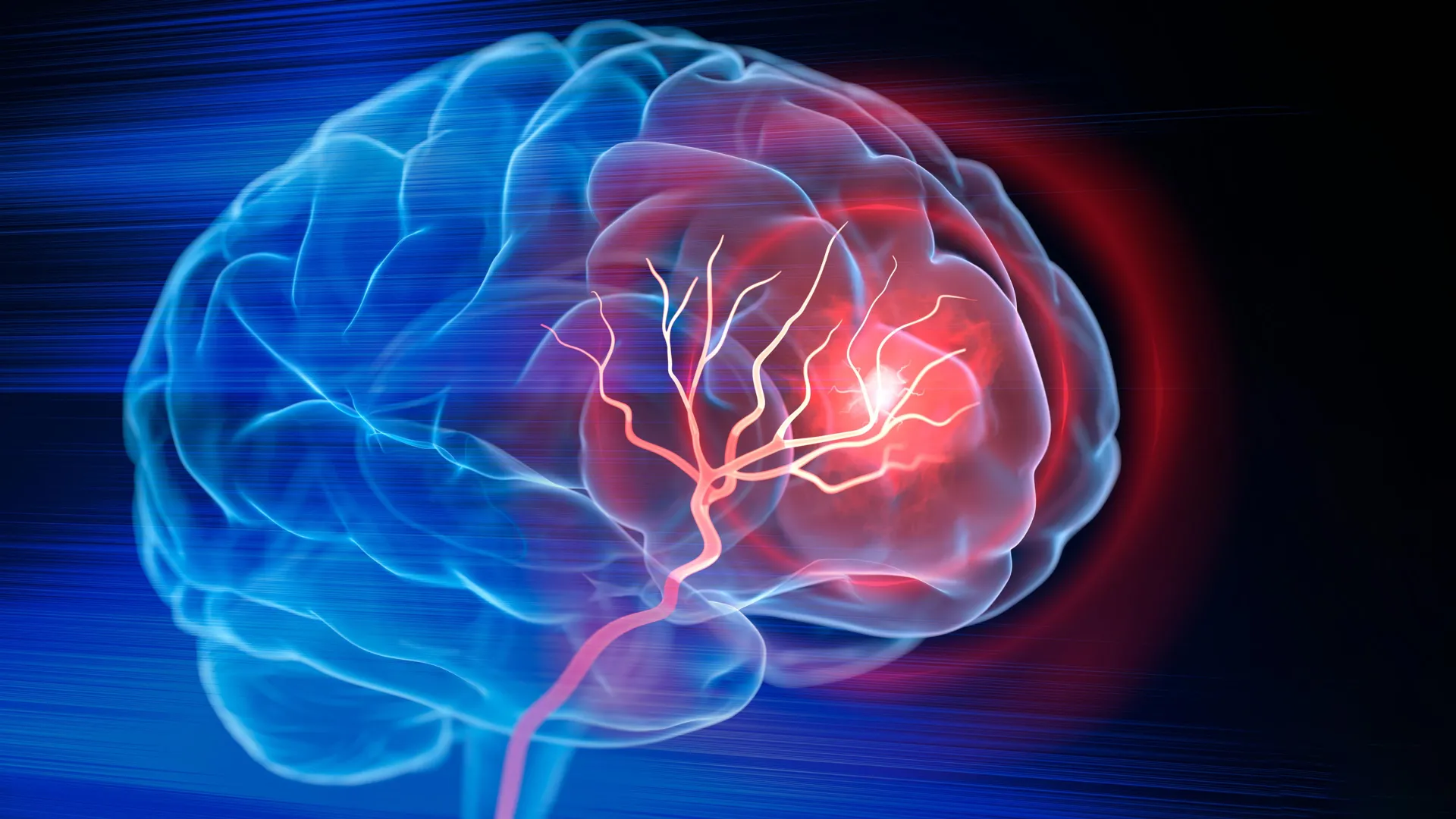
An injectable biomaterial helped mice recover movement after ischemic stroke by creating a local environment that supported blood-vessel formation, neural growth and immune-cell activity, according to research from Duke University. The findings, published in Cell Biomaterials, remain preclinical and have not established that the approach works in people.
The Duke team used microporous annealed particle scaffolds, or MAPS. Made from hydrogel microparticles, the material forms an open structure that cells can enter. Researchers injected the scaffold directly into the cavity left by brain tissue destroyed during a stroke.
To enhance the scaffold, the researchers attached extracellular vesicles derived from laboratory-grown astrocytes to the microparticles. Astrocytes are support cells in the brain, while extracellular vesicles are microscopic packages containing proteins, lipids and genetic material that can influence other cells. The design was intended to keep these signals concentrated within the damaged region rather than allowing them to disperse.
A combination of the signaling molecules IL-4 and C1q was especially effective at attracting immune cells to the injury site. These included macrophages and a persistent population of neutrophils. Neutrophils are commonly associated with inflammation and tissue damage soon after stroke, but the study suggested that their role may differ later in recovery depending on their surroundings and the signals they receive.
When researchers reduced the neutrophil-rich immune-cell population, the treated tissue produced substantially fewer blood vessels and the scaffold underwent less remodeling. This result indicated that the recruited cells contributed to the repair response observed in the mice.
The biological changes were accompanied by functional improvement. Treated animals developed more axonal fibers in and around the damaged region and performed better on a grid-walking test measuring forelimb-placement errors. After eight weeks, their performance was not statistically distinguishable from that of healthy control mice, and the improvement continued for the rest of the study.
The extracellular vesicles did not produce comparable blood-vessel repair when administered without the MAPS scaffold. That comparison suggested that the material’s porous architecture and ability to retain the vesicle-associated signals were important parts of the response, rather than the vesicles acting alone.
The researchers emphasized that the treatment is still at an early stage. The experiments involved mouse models and direct injection into the damaged brain, so questions about safety, immune-cell behavior and performance in larger models remain unresolved. The team is also investigating extracellular vesicles produced by human induced pluripotent stem cell-derived astrocytes, which could provide a more scalable and clinically relevant source for future research.
Reporting Note
Biohack Report distinguishes preliminary findings, clinical evidence and commercial claims whenever the available reporting supports that distinction. Coverage is informational and is not medical advice.