Mouse Study Identifies Brain Circuit That Compares New Sights With Recent History
MIT researchers identified a thalamo-cortical pathway in mice that helps the frontal cortex interpret current visual evidence in light of what happened moments earlier. The circuit influenced whether animals maintained or changed decisions in a moving-dots task, though the findings remain preclinical.
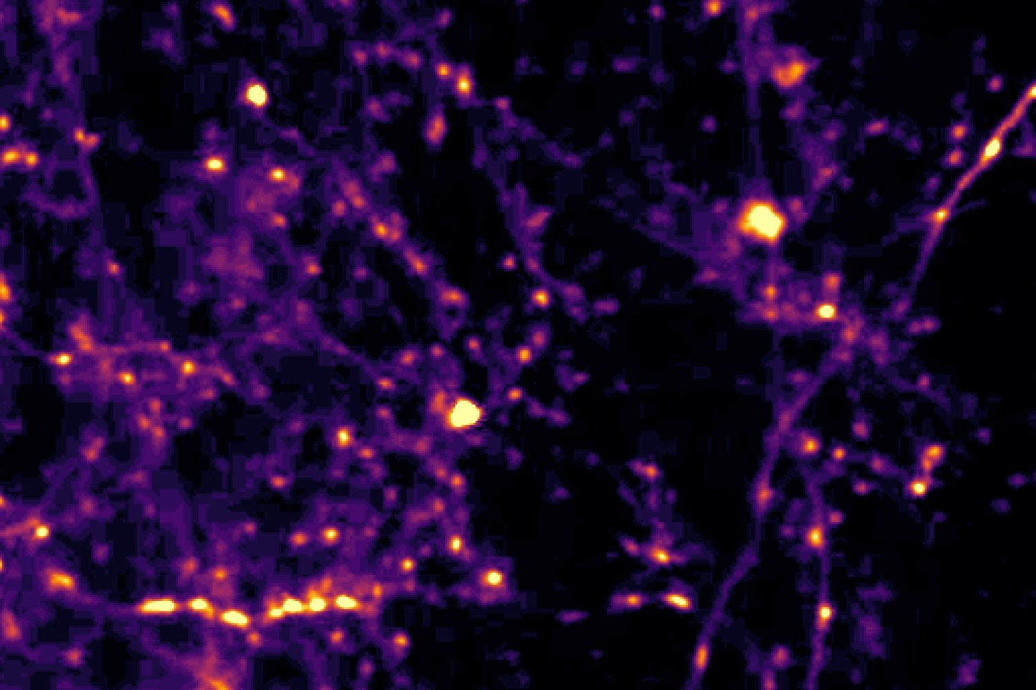
A brain circuit in mice helps compare current visual information with what was seen moments earlier, according to a study published in Science by researchers at MIT’s Picower Institute for Learning and Memory. The pathway appeared to influence whether the animals maintained an existing decision or reconsidered it when sensory evidence changed.
The circuit connects the lateral posterior thalamus, or LP, with the anterior cingulate cortex, or ACC. The researchers describe the LP as providing the frontal cortex with information about how much the current sensory cue differs from the immediate past, while the ACC uses that comparison in forming a choice. In humans, the corresponding thalamic region is called the pulvinar, and the researchers said the circuitry is closely analogous to systems involved in sensory decisions in the human prefrontal cortex.
To study the process, the team trained mice to play a visual task involving dots drifting across a screen. Some portion of the dots moved together either to the left or right, and the mice had to identify the overall direction. The proportion of coherently moving dots varied between trials, allowing the researchers to create different degrees of similarity or difference between consecutive cues.
The animals’ choices showed that recent sensory history affected their behavior. When a mouse had made a correct guess and the next cue was similar, it was likely to repeat that choice. When the new cue differed substantially, repeating the choice became less likely. After an incorrect guess, the pattern reversed: similar cues made the mouse less likely to repeat the decision, while markedly different cues made repetition more likely.
The researchers used two-photon microscopy to record activity in the LP-ACC pathway and neurons in the ACC while the mice performed the task. They also used optogenetics to artificially perturb inputs from the LP into the ACC. Stimulating the pathway in one hemisphere made mice less likely to choose rightward motion, while stimulation in the other made rightward choices more likely. In both cases, the behavioral disruption depended on how different the current cue was from the preceding one.
The findings indicate that the pathway was causally involved in how the mice used recent sensory information, rather than simply carrying signals unrelated to the decision. The researchers reported that the LP and ACC performed distinct roles: the LP supplied a history-referenced comparison, while the ACC translated that information into activity associated with the animal’s eventual choice.
The work was conducted in mice and does not establish that the same circuit produces identical effects in humans. MIT researchers said the findings may be relevant to research on autism, where differences in sensory prediction and filtering have been reported, but the study did not test an autism intervention or demonstrate a clinical application.
Reporting Note
Biohack Report distinguishes preliminary findings, clinical evidence and commercial claims whenever the available reporting supports that distinction. Coverage is informational and is not medical advice.